
Bacteria
Structure
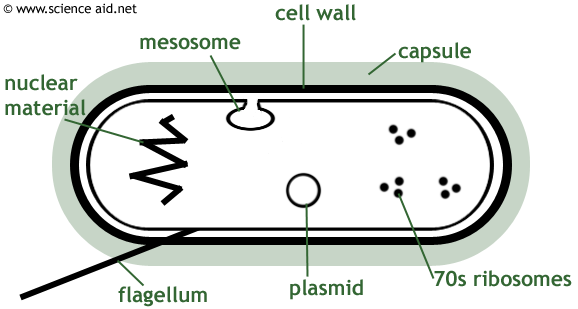
The following are the functions of the various structures of the various features of the bacterial cell.
| Cell Wall | Protects the cell and maintains its shape, bacteria can be catagorized according to their cell wall type: Gram positive walls are thick with little lipid. Gram negative walls are much thinner, with two layers. |
| Capsule | Some bacteria have a slimy layer of polysaccharides and polypeptides, allowing them to attach to objects and providing protection. |
| 70s Ribosomes | These are for performing protein synthesis. Are smaller than the 80s ribosomes in eukaryotic cells |
| Flagellum | Another optional feature, is a projection that moves around to allow the cell to move. A cell may have multiple flagella arranged around it. |
| Nuclear Material | A folded mass of DNA and RNA, also called the nuclear zone - containing all the genes required for vital functions. |
| Plasmid | Plasmids are additional rings of genetic material that aren't essential to the cell, often contains genes for antibiotic resistance. |
| Mesosome | An infolding of the membrane that is the site of respiration (like a mitochondrion) - it's shape improves surface area. The existance of the mesosome is disputed, and most scientists believe it is a mistake in electron microscope technique. |
Bacterial Growth
Bacteria are very flexible and obtain energy in a number of ways. This includes respiration at the mesosome; many bacteria perform photosythesis at additional sites with specialized pigments. Energy can also be obtained by oxidising inorganic compounds, using the energy to synthesize organic compounds - nitrifying bacteria do this.
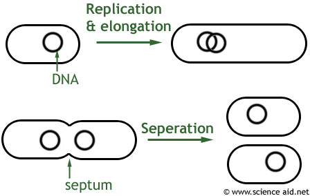
Bacteria reproduce by an asexual process called binary fission. First, the DNA replicates and the cell elongates. In the middle of the elongated cell, a septum forms and this develops in to a cell wall that divides two seperate cells.
The population of bacteria follows a predictable pattern, and this can be represented as follows.
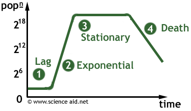
The first phase is the lag phase the cells are active but do not increase very much. The exponential phase follows where a plentiful supply of nutrient and space allow an ever-increasing rate of growth, and bacterial production outstrips bacterial death.
Once the carrying capacity (the maximum population the environment can support), is reached, the population enters the stationary phase where no net change in population occurs. The environment is changed by the bacteria as metabolic waste builds up and the conditions become increasingly difficult. This leads to the death (or final) phase where more cells die that are produced, as a result of waste toxicity, starvation and oxygen shortage; and the population declines.
Culturing and Counting Bacteria
When growing bacteria in the lab, it is important to use aseptic conditions that will prevent contamination by other micro-organisms and protect scientists from growing pathogens.
Techniques used include heating instruments (innoculating loop for example) over a bunsen burner; flaming the neck of tubes; and opening the Petri dish lid as little as possible to reduce contamination from air-borne micro-organisms.
When culturing bacteria it is often necessary to count how many bacteria there are. There are a variety of methods used to determine this.
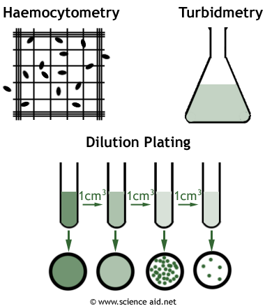
Haemocytometry is a total count method where every cell (dead or alive) is counted. It works by introducing a standard amount of bacteria solution in to the haemocytometer - a glass slide with lots of grid lines. This is placed under a microscope and the number of cells counted using a standard method.
A far simpler technique is turbidmetry which works on the principle that a more turbid (cloudy) solution has more cells. The light absorbed is recorded and can be compared with reference graphs to estimate the number of cells. This is also a total cell count method.
In dilution plating, 1cm3 of original bacterial solution is taken and diluted with 9cm3 of water. The same is done with this diluted solution and so on. A sample from each dilution is cultured; once individual colonies can be seen, it means each of those represents a single bacterium that was in the solution. The number is multiplied by the dilution factor.
This is a viable cells count method - meaning it only counts those bacteria which are alive, since dead bacteria can not grow colonies. Depending on the aim of your investigation, this could be a much more useful type of count.
When a bacterial solution is spread out evenly over an agar surface, it will produce a bacterial lawn. It is used in testing the effectiveness of antibiotics or disinfectants.

B has a zone of inhibition which means it has prevented bacteria growing. A, however, has had no affect. The size of the zone is indicative of the substance's concentration, rather than it's effectiveness.